
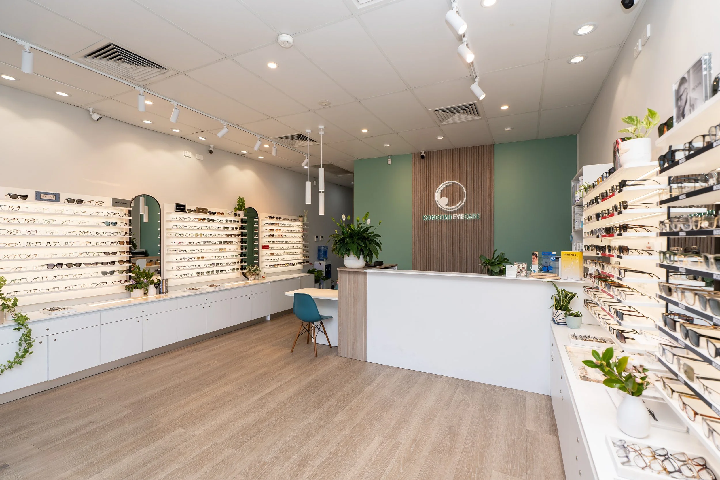
About Concord Eyecare - Independent Optometrists in North Strathfield & The Inner West
We’ve been looking after inner west eyes for over 20 years.
We're a family-run independent practice — not a chain, not a franchise, not a corporate clinic with rotating staff. Just a small team of experienced optometrists who genuinely care about getting it right.
★ 4.9 Google Rating · 20+ Years · 3 Optometrists · All Health Funds Accepted
Our Story: 20 years of independent optometry in the Inner West
Twenty years of doing things the right way.
Concord Eyecare has been part of the inner west for over two decades. We believe in taking the time to listen properly, explain everything in plain language, and make sure you leave understanding your eyes — not just your prescription.
What has changed is the technology. We've invested in advanced diagnostic equipment like OCT scanning and corneal topography. We've built a dedicated myopia control clinic for children. We offer IPL therapy for chronic dry eye. And all three of our optometrists are therapeutically endorsed — meaning we can prescribe medications directly, without a GP referral.
We're independently owned, which means we choose our own equipment, our own lens suppliers, and our own frame brands based on what's actually best — not what head office tells us to sell. The optometrist you see today is the same one who'll remember your history next year.
That's what independent optometry should feel like.
WHAT MAKES CONCORD EYECARE DIFFERENT FROM OTHER OPTOMETRISTS
A few things we do that makes us different
We don't rush appointments
No conveyor belt here. We allocate enough time to be thorough — 30 minutes minimum, longer if you need it.
We can prescribe directly
All three optometrists are therapeutically endorsed — we can prescribe medications for eye infections, allergies, glaucoma, and dry eye without a GP referral.
We're independently owned
No head office, no sales targets, no corporate lens deals. We recommend what's best for your eyes — from whichever brand that happens to be.
Paediatric training across the team
All three optometrists have advanced paediatric qualifications. Children's vision isn't a sideline for us — it's a core focus.
Advanced diagnostic equipment
OCT scanning, corneal topography, retinal imaging, and axial length measurement. We invest in the equipment that lets us catch problems early.
Continuity of care
You'll see the same optometrist every visit. We know your history, your family, and your eyes — no starting from scratch each time.
SERVICES WE HAVE SPECIAL INTEREST IN
At Concord Eyecare we cover the full range of clinical optometry —
from comprehensive eye examinations, children's eye care, myopia control and ortho-k, to advanced dry eye treatment including IPL therapy and meibomian gland dysfunction management. Each of us has areas of clinical interest we've trained deeply in, which lets us offer genuinely evidence-based care across the board.
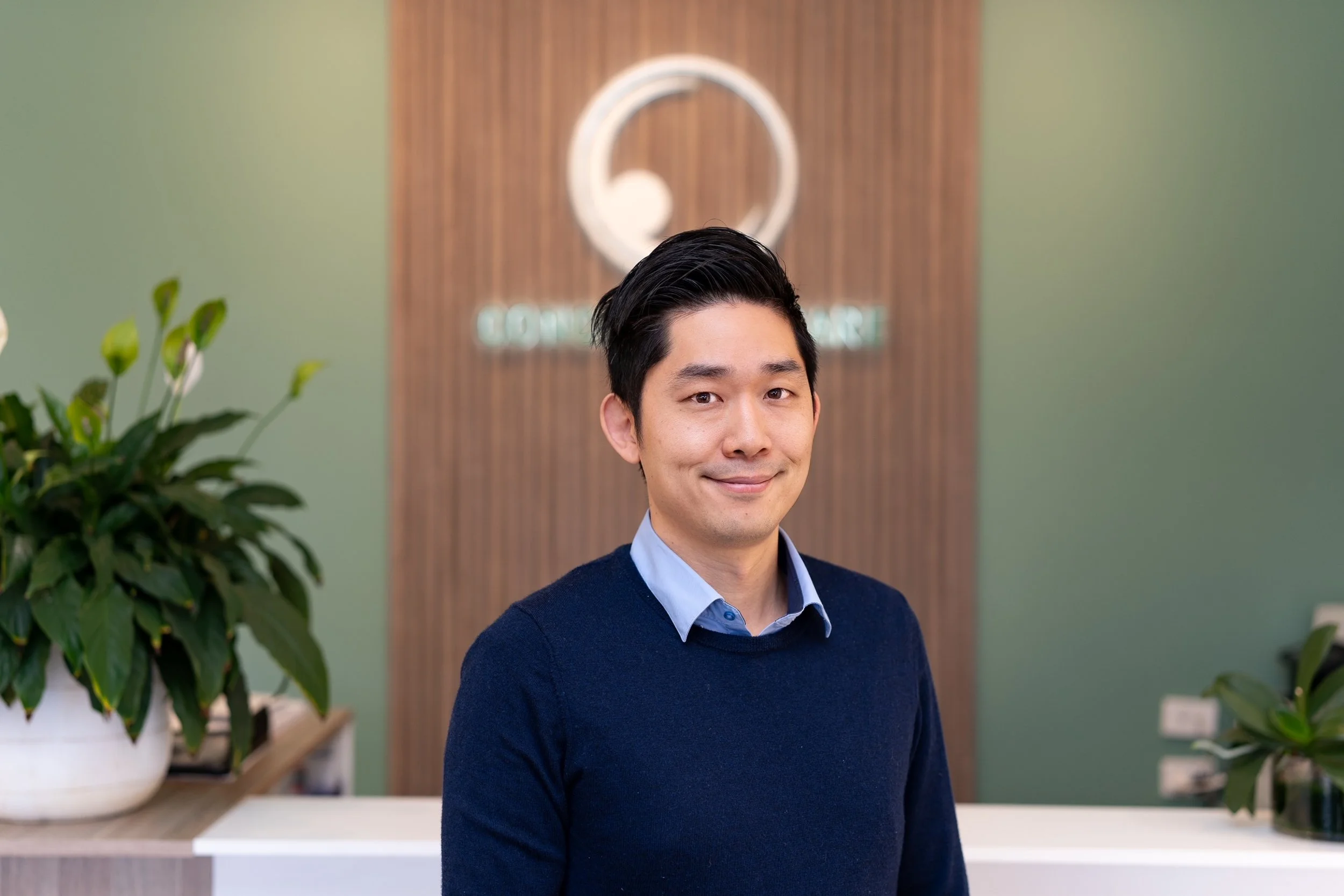
Dr Mark Joung
Principal Optometrist
Mark graduated from UNSW in 2006 with Honours in Optometry and the Ciba Vision Award for contact lens performance. He's therapeutically endorsed — meaning he can prescribe medications directly without a GP referral — and holds postgraduate qualifications in Advanced Paediatric Eye Care (UNSW) and Ocular Therapeutics.
He has a special interest in myopia control, orthokeratology, and dry eye management — partly because he's a high myope himself, so he genuinely understands what his patients are going through. He's a member of the Orthokeratology Society of Oceania and has worked closely with ophthalmologists in managing glaucoma, diabetes, and complex eye conditions.
Outside the practice, Mark is a dad of two who spends his downtime cycling, gardening, and being dragged to playgrounds.
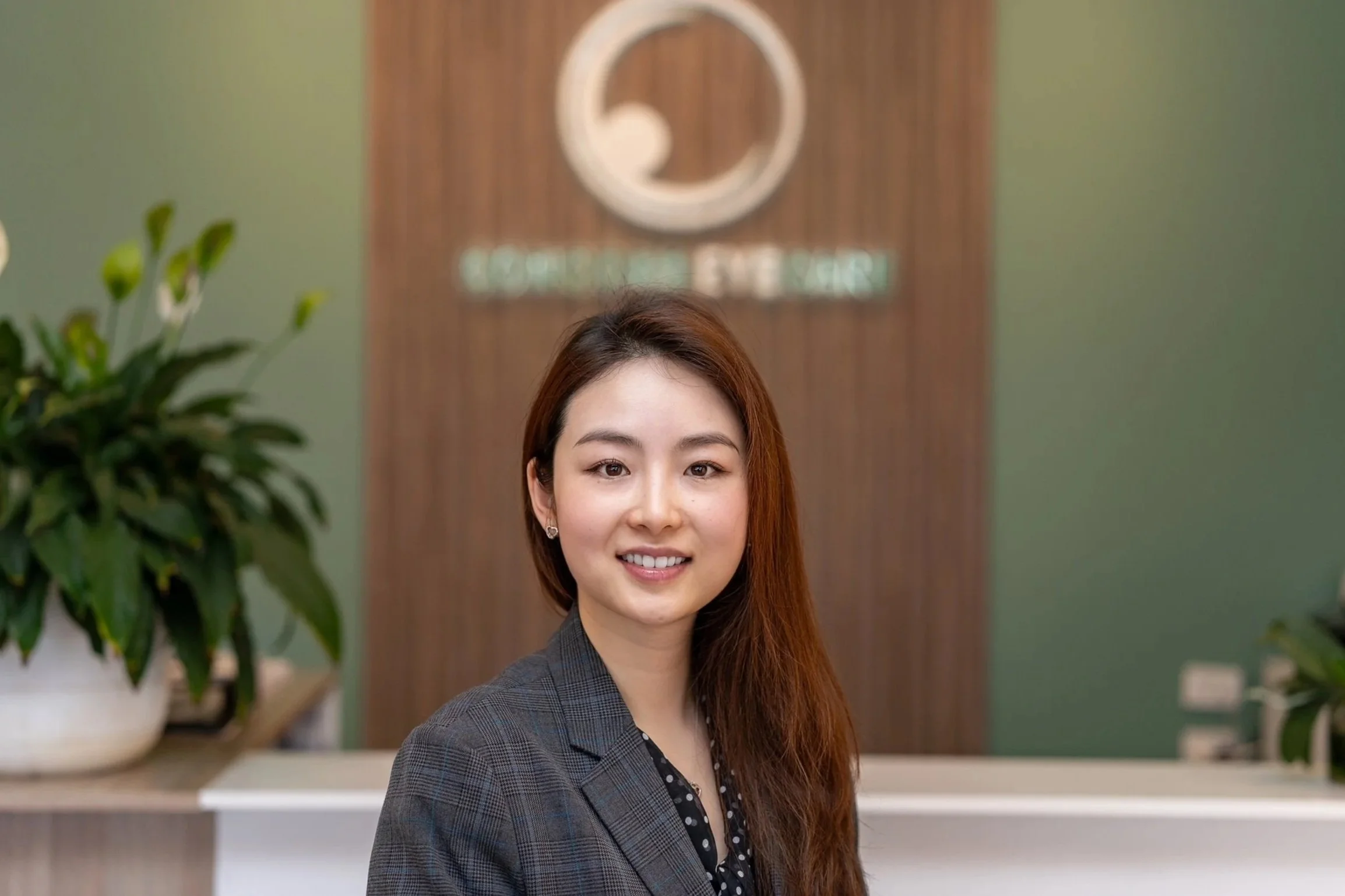
Dr Nikki Peng
OPTOMETRIST
Nikki graduated from UNSW in 2006 with First Class Honours and spent over five years in contact lens research at the Brien Holden Vision Institute — one of the world's leading eye research centres. That research background shows in the way she approaches fittings.
She's therapeutically endorsed with a Graduate Certificate in Ocular Therapeutics from UNSW, and holds an Advanced Children's Vision qualification from the Australian College of Optometry. At Concord Eyecare she focuses on children's vision and myopia control, dry eye disease, and ocular disease co-management, working closely with local ophthalmologists.
Her published work in mivision on dry eye disease reflects a long-standing clinical interest that continues at the practice today.
When she's not in the practice, Nikki is a mum of two who enjoys creative arts in her spare time.
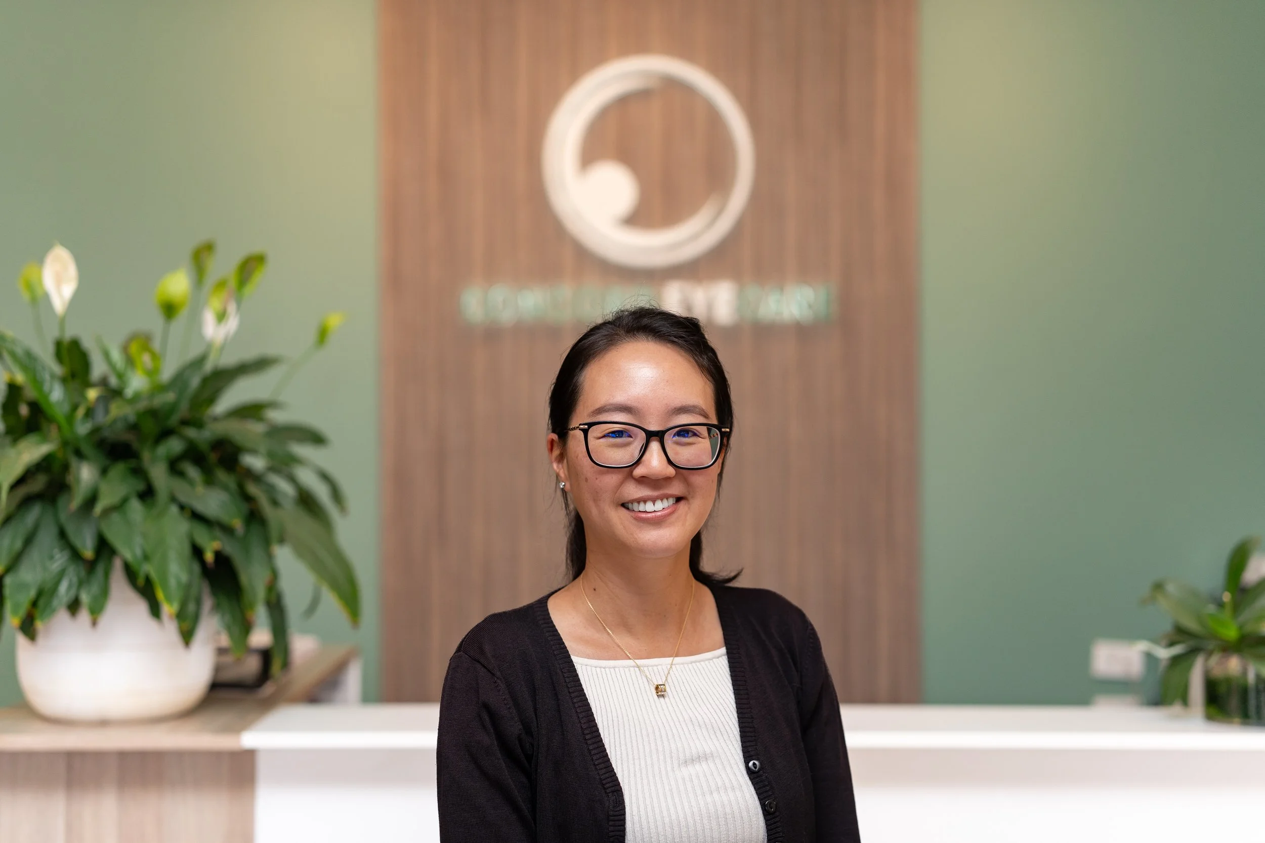
Dr Vivian Li
OPTOMETRIST
Vivian graduated with Honours in Optometry in 2004 and was awarded the Australasian College of Behavioural Optometrist Prize for best overall performance in Binocular and Children's Vision — which gives you a sense of where her passion lies.
She's therapeutically endorsed with a Graduate Certificate in Ocular Therapeutics, and holds an Advanced Children's Vision qualification from the Australian College of Optometry. She has a special interest in children's vision and myopia management, and brings extensive experience in ocular disease co-management from years working alongside GPs and ophthalmologists — including time in rural NSW in Dubbo and Cobar, where you learn to handle just about everything.
The Diagnostic equipment we use
We invest in the equipment that matters.
We don't just rely on the basics. Our practice is equipped with advanced diagnostic technology that lets us see problems earlier and more accurately than a standard eye test.
-
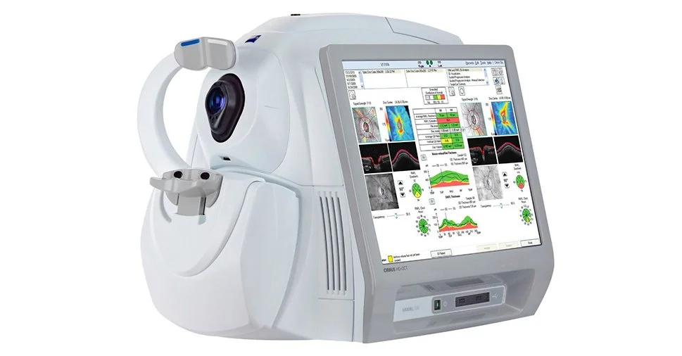
![image of a Carl Zeiss OCT]()
OCT scanner
Cross-section imaging of your retina and optic nerve. Picks up glaucoma and macular changes years before symptoms appear.
-
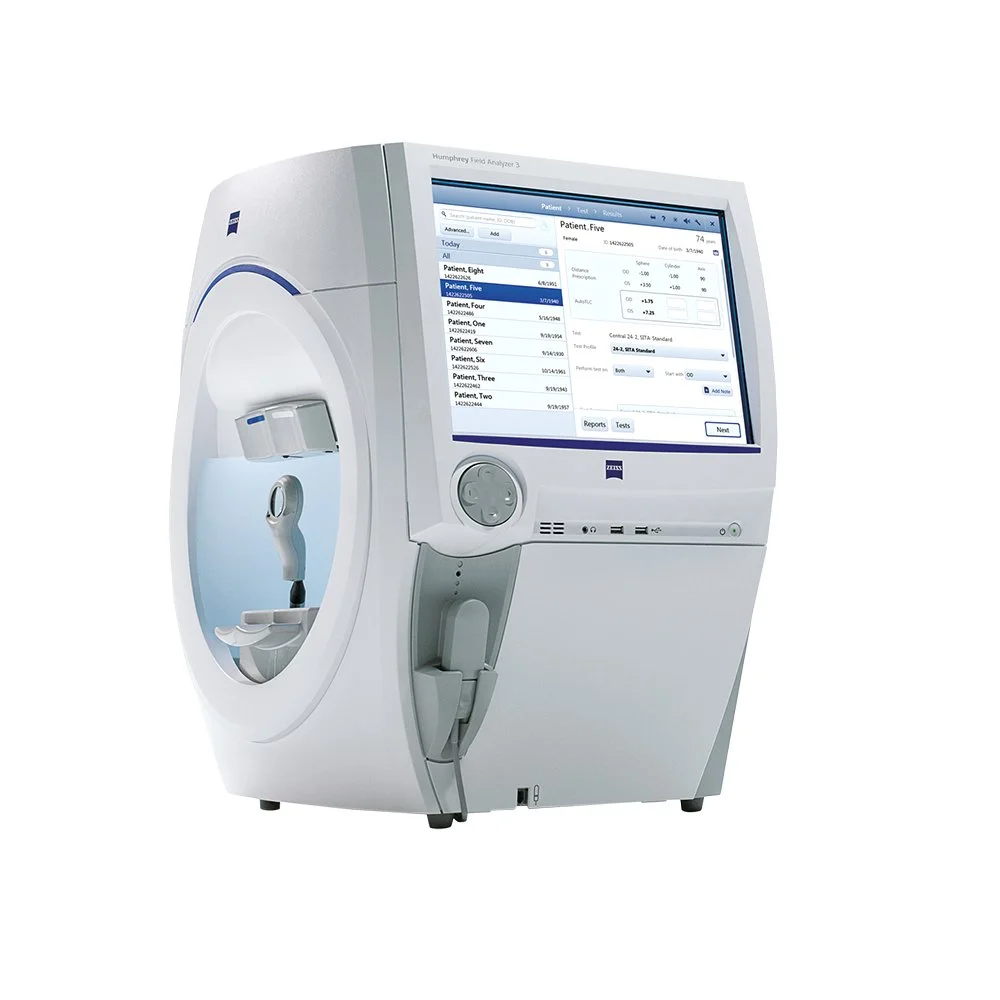
![image of Carl Zeiss HFA3]()
Visual Field Analyser
Tests peripheral vision — critical for glaucoma, neurological conditions, and driving fitness assessments.
-
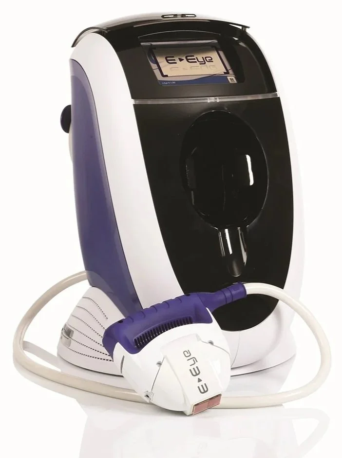
![image of e-eye IPL]()
E-Eye IPL
Intense pulsed light therapy for meibomian gland dysfunction — the most common cause of chronic dry eye.
-
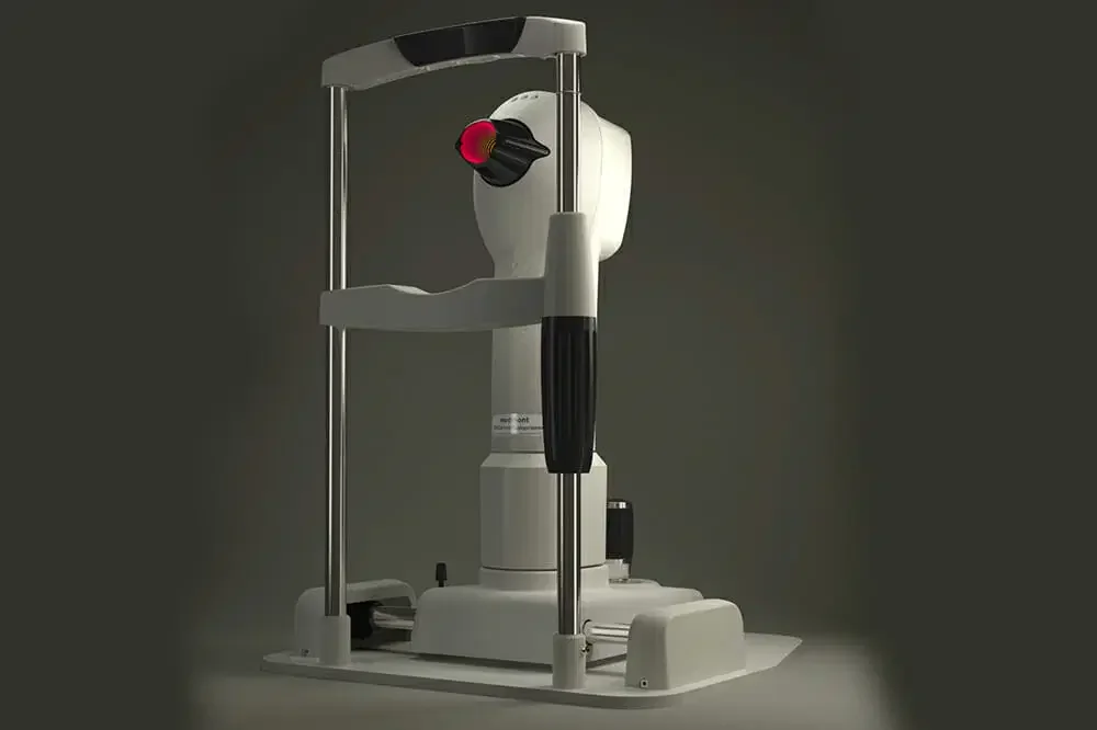
![image of Medmont E300 corneal topographer]()
Corneal Topographer
Maps the surface of your cornea in detail. Essential for contact lens fitting, Ortho-K, and detecting keratoconus.
THE Concord Myopia Clinic- Children’s Myopia Control
We also run a dedicated myopia control clinic.
Myopia in children is something we take seriously — seriously enough to build a whole clinic around it. The Concord Myopia Clinic runs alongside our general practice and offers Ortho-K, MiSight, MiyoSmart, and atropine treatment with transparent pricing and a team that's been fitting these lenses longer than most.
Visit us in North Strathfield
Come and meet us
No referral needed. We’re open Monday to Saturday at 161 Concord Rd North Strathfield. Book online or give us a call.